Mural paintings
Caroline Bleeker produces the phase contrast microscope
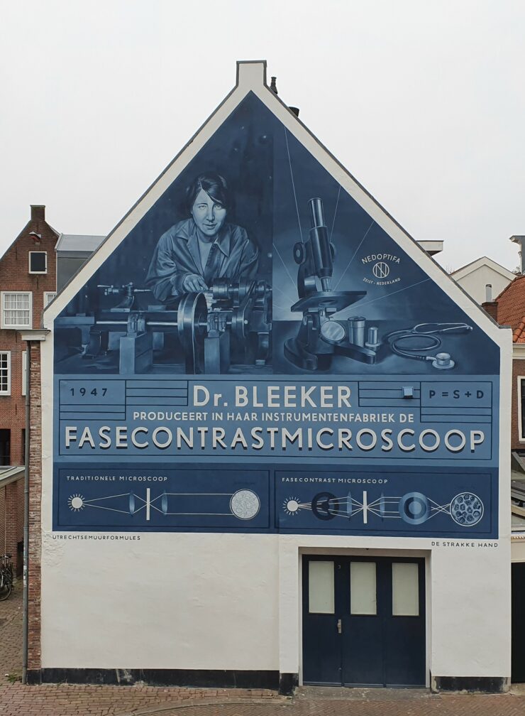
What do we see here?
 Scientific research requires accurate instruments, such as microscopes. These instruments are often developed by scientists and instrument makers at universities. Caroline Bleeker developed the phase contrast microscope together with University of Groningen Professor and Nobel Prize winner Frits Zernike. They received a patent for their invention in 1947.
The phase contrast microscope makes it possible to see the interior structure of living cells, allowing researchers to follow the process of cell division in living bacteria, for example. Before the invention of the phase contrast microscope, we could only study living cells by using dyes. One of the major disadvantages of cell dyes is that they can kill the cells. Phase contrast technology was therefore vital for the fields of biology and medicine, and is still widely used today in hospitals, businesses and universities.
The story of the discovery and development of the phase contrast microscope is a wonderful example of how research in a particular field – in this case theoretical physics – can make an important contribution to completely different fields of research, such as biology and medicine.
Caroline Bleeker set up a company called NEDOPTIFA, which produced large numbers of phase contrast microscopes.
Frits Zernike came up with a way to enhance the contrast for extremely thin samples, such as single cells. His idea is based on the fact that light behaves like a wave. The more light, the higher the wave. When the light passes through a very thin sample, such as a single cell, the intensity is only slightly diminished and there is almost no difference between the light that doesn’t pass through the cell. So it’s hard to see the difference between the cell and the background. You can see this effect in the illustration at the bottom right, taken with a traditional microscope.

Cells from an onion captured by a phase-contrast microscope (left) and a traditional microscope (right). Wikipedia, Creative Commons Attribution-Share Alike 4.0 International license.
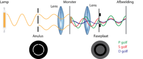
The light passing through the sample is also slightly delayed compared to the light passing around the cell. In physics, we call this phenomenon ‘phase shift’. We can label the two light waves with the letters ‘S’ (the original light wave) and ‘D’ (the light wave shifted by the sample). Mixing these two waves together creates a new wave, called ‘P’. Like waves of water, light waves can amplify and interfere with one another. This amplification and interference of the waves creates higher and lower waves, and therefore stronger contrasts in the final image.
To separate the light waves ‘S’ and ‘D’ and re-mix them into ‘P’, the phase contrast microscope has some additional components that a traditional microscope does not have: the annular diaphragm and the phase plate. The figure below shows how the waves move through a phase-contrast microscope.

Waves in a phase-contrast microscope. The ‘S’, ‘D’ and ‘P’ waves are shown in red, blue and green.
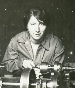
 Caroline Bleeker (1897 – 1985) earned her PhD in Physics cum laude at Utrecht University in 1928. Her supervisor was Prof Leonard Ornstein. Less than two years later, she founded a consulting bureau at Korte Nieuwstraat 13, just a stone’s throw from the Strosteeg in Utrecht. Her ‘Physisch Adviesbureau’ consulted on scientific instrumentation. That same year, Caroline Bleeker opened an instrument factory in Utrecht as well. Her successful business produced scientific instruments for laboratories, and later the company added a department for optical instruments as well. During the Second World War, Caroline Bleeker hid Jewish fugitives in her factory, and received a royal honour for her resistance work after the war.
Caroline Bleeker also advocated for women’s emancipation. She was an active female scientist in a time when that was still a very rare achievement. Founding her own company also made her an independent entrepreneur, and she hired both male and female employees to work in her factory. The company magazine at NEDOPTIFA even had a special column for women.
Caroline Bleeker also advocated for women’s emancipation. She was an active female scientist in a time when that was still a very rare achievement. Founding her own company also made her an independent entrepreneur, and she hired both male and female employees to work in her factory. The company magazine at NEDOPTIFA even had a special column for women.
Where can you find this mural painting?

